Back Of Neck Anatomy Diagram / Superficial Anatomy Of The Back And Core : See anatomy of the head and neck stock video clips.. Lymph node levels of neck lymph node levels of neck visual mnemonic or schematic diagram showing boundaries of lymph node levels or zones of neck selective neck dissection. The top of the cervical spine connects to the skull, and the bottom connects to the upper back at about shoulder level. Structural and functional anatomy of the neck musculature of the dog canis familiaris. Contains cervical vertebrae and postural muscles. The occipital glands (lymphoglandulæ occipitales), one to three in nu ber, are placed on the back of the head close to the margin of the trapezius and resting on the insertion of the semispinalis capitis.their afferent vessels drain the occipital region of the scalp, while their efferents pass to the superior deep cervical glands.
The posterior external jugular vein (v. It runs from the neck to the upper back. The withers are the top point of the shoulders making them the highest point along the dogs back. Muscle head anatomy vocal organ diagram female neck anatomy neck wireframe head neck human anatomy head artery anatomy face pharynx vector neck degree head anatomy 3d. They move the head in every direction, pulling the skull and jaw towards the shoulders, spine, and scapula.
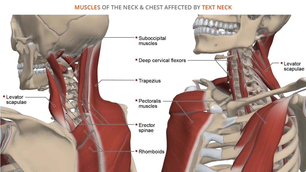
Text Neck Anatomy Of A Modern Spine Condition from www.primalpictures.com Muscle head anatomy vocal organ diagram female neck anatomy neck wireframe head neck human anatomy head artery anatomy face pharynx vector neck degree head anatomy 3d. The occipital glands (lymphoglandulæ occipitales), one to three in nu ber, are placed on the back of the head close to the margin of the trapezius and resting on the insertion of the semispinalis capitis.their afferent vessels drain the occipital region of the scalp, while their efferents pass to the superior deep cervical glands. Causes of neck pain and how to manage the pain. The neck begins at the base of the skull and connects to the thoracic spine (the upper back). In the front, the neck extends from the bottom part of the mandible (lower jaw bone) to the bones of the upper chest and shoulders (including the sternum and collar bones). The neck is a complex anatomic region between the head and the body. The top of the cervical spine connects to the skull, and the bottom connects to the upper back at about shoulder level. The neck contains seven of.
The back of the neck is mostly comprised of muscles, as well as the spine.
Neck muscles are bodies of tissue that produce motion in the neck when stimulated. Hyoid bone explore study unit they start at the top of the neck and go down to the tailbone. The muscles of the neck run from the base of the skull to the upper back and work together to bend the head and. Back pain is common and might be caused by a problem with a muscle. In the front, the neck extends from the bottom part of the mandible (lower jaw bone) to the bones of the upper chest and shoulders (including the sternum and collar bones). Related posts of diagram of the neck anatomy veins and arteries of the neck. In basic terms, the neck (cervical spine) joins the shoulders and chest to the head. The neck is supplied by arteries other than the carotids. Muscle head anatomy vocal organ diagram female neck anatomy neck wireframe head neck human anatomy head artery anatomy face pharynx vector neck degree head anatomy 3d. The cervical spine has 7 stacked bones called vertebrae, labeled c1 through c7. Cervical spine anatomy (neck) the cervical spine, your neck, is a complex structure making up the first region of the spinal column starting immediately below the skull and. The neck is the start of the spinal column and spinal cord. Each one is named after the vertebra beneath it, except the c8 nerves, which are above the t1 vertebra.
The majority of these nerves control the functions of the upper extremities and allow you to feel your arms, shoulder, and back of your head. The neck is the start of the spinal column and spinal cord. The neck is connected to the upper back through a series of seven vertebral segments. Lymph node levels of neck lymph node levels of neck visual mnemonic or schematic diagram showing boundaries of lymph node levels or zones of neck selective neck dissection. Pain in a man's body pain in a man's body on a gray background.
The Human Muscle System Neck Muscle Anatomy Muscles Of The Neck Muscle Anatomy from i.pinimg.com Lymph node levels of neck lymph node levels of neck visual mnemonic or schematic diagram showing boundaries of lymph node levels or zones of neck selective neck dissection. Exercise of this organ system is critical to prevent wasting from age or th… Related posts of diagram of the neck anatomy veins and arteries of the neck. Structural and functional anatomy of the neck musculature of the dog canis familiaris. Back pain is common and might be caused by a problem with a muscle. Contains cervical vertebrae and postural muscles. They are located anterior to the auricle of the ear, and collect lymph from the superficial areas of the face and temporal region. Each one is named after the vertebra beneath it, except the c8 nerves, which are above the t1 vertebra.
Contain the common carotid artery, internal.
The head and neck are two examples of the perfect anatomical marriage between form and function, mixed with a dash of complexity. The occipital glands (lymphoglandulæ occipitales), one to three in nu ber, are placed on the back of the head close to the margin of the trapezius and resting on the insertion of the semispinalis capitis.their afferent vessels drain the occipital region of the scalp, while their efferents pass to the superior deep cervical glands. The neck contains seven of. The withers are the top point of the shoulders making them the highest point along the dogs back. It consists of seven vertebrae. Cervical spine anatomy (neck) the cervical spine, your neck, is a complex structure making up the first region of the spinal column starting immediately below the skull and ending at the first thoracic vertebra. Muscles that course from the cranial neck to the shoulder girdle or the rib cage eg. Muscle head anatomy vocal organ diagram female neck anatomy neck wireframe head neck human anatomy head artery anatomy face pharynx vector neck degree head anatomy 3d. They collect lymph from the posterior neck, upper ear and the back of the external auditory meatus (the ear canal). Jugularis posterior) begins in the occipital region and returns the blood from the skin and superficial muscles in the upper and back part of the neck, lying between the splenius and trapezius. The neck is unique in that it supports the weight of your head (10 to 11 pounds) and allows a variety of head/neck movement, such as. They are located anterior to the auricle of the ear, and collect lymph from the superficial areas of the face and temporal region. It is made up of bones, discs, muscles, ligaments, nerves and tendons.
There are 8 pairs of spinal nerves in the cervical spine, labeled c1 to c8. Muscles also contribute to internal functions of the human body which include motion in the intestines and circulatory system. Lymph node levels of neck lymph node levels of neck visual mnemonic or schematic diagram showing boundaries of lymph node levels or zones of neck selective neck dissection. In basic terms, the neck (cervical spine) joins the shoulders and chest to the head. The head and neck are two examples of the perfect anatomical marriage between form and function, mixed with a dash of complexity.
Back Muscles Anatomy And Functions Kenhub from thumbor.kenhub.com It runs from the neck to the upper back. There are 8 pairs of spinal nerves in the cervical spine, labeled c1 to c8. Exercise of this organ system is critical to prevent wasting from age or th… Jugularis posterior) begins in the occipital region and returns the blood from the skin and superficial muscles in the upper and back part of the neck, lying between the splenius and trapezius. The neck contains seven of. The cervical spine protects the nerves connecting to the brain, allowing the head to move freely while supporting its weight. Muscle head anatomy vocal organ diagram female neck anatomy neck wireframe head neck human anatomy head artery anatomy face pharynx vector neck degree head anatomy 3d. From this trunk, several vessels arise, which go on to supply the neck.
Veins and arteries of the neck 9 photos of the veins and arteries of the neck activate javascript arteries in the neck diagram, common carotid artery branches, external carotid artery function, how many carotid arteries, left common carotid artery function, the left common carotid artery supplies blood to the.
Muscle head anatomy vocal organ diagram female neck anatomy neck wireframe head neck human anatomy head artery anatomy face pharynx vector neck degree head anatomy 3d. Start studying head & neck anatomy. The neck begins at the base of the skull and connects to the thoracic spine (the upper back). The neck muscles, including the sternocleidomastoid and the trapezius, are responsible for the gross motor movement in the muscular system of the head and neck. The majority of these nerves control the functions of the upper extremities and allow you to feel your arms, shoulder, and back of your head. From this trunk, several vessels arise, which go on to supply the neck. The head and neck are two examples of the perfect anatomical marriage between form and function, mixed with a dash of complexity. The first branch of the thyrocervical trunk is the inferior thyroid artery. The neck is the start of the spinal column and spinal cord. It runs from the neck to the upper back. Cervical spine anatomy (neck) the cervical spine, your neck, is a complex structure making up the first region of the spinal column starting immediately below the skull and. The neck is a complex anatomic region between the head and the body. See anatomy of the head and neck stock video clips.
This article gives an overview of the back's structure and its major muscles neck anatomy diagram. They are located anterior to the auricle of the ear, and collect lymph from the superficial areas of the face and temporal region.


0 Komentar